
Clinical resources · Pancreatobiliary
EUS and ERCP learning resources
Explore the existing clinical summaries, procedural anatomy, regional pathways and training resources.
Management of pancreatic cystic lesions (North East regional pathway, 2026)
Scope and currency. This pathway was created in 2026 and applies to the North East region only. It is not a national guideline. Before using it, confirm you are looking at the current version, or follow your own local pathway. If in doubt, discuss with the regional MDT.
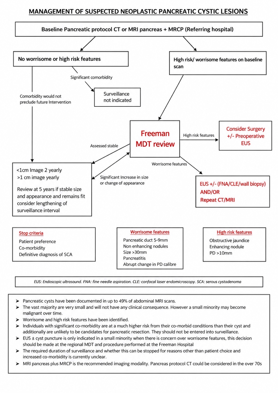
Worrisome features
- Pancreatic duct 5-9mm
- Non enhancing nodules
- Size >30mm
- Pancreatitis
- Abrupt change in PD calibre
High risk features
- Obstructive jaundice
- Enhancing nodule
- PD >10mm
Stop criteria
- Patient preference
- Co-morbidity
- Definitive diagnosis of SCA
Surveillance interval
- <1cm image 2 yearly
- >1cm image yearly
- Review at 5 years. If stable size and appearance and remains fit, consider lengthening of surveillance interval
Significant co-morbidity that would preclude future intervention: surveillance not indicated.
EUS: endoscopic ultrasound. FNA: fine needle aspiration. CLE: confocal laser endomicroscopy. SCA: serous cystadenoma. PD: pancreatic duct.
ERCP
Endoscopic retrograde cholangiopancreatography (ERCP) uses a flexible side-viewing duodenoscope to reach the ampulla of Vater (simply called ampulla) under both endoscopic & fluoroscopic guidance. This contrasts to a forward viewing scope used in gastroscopy – the ERCP scope is akin to driving a car with the view as you are looking of a side side window whilst driving forwards.This means the operator must have a higher level of understanding of fine tip control ( as intubation is often done blindly through feel) and of the anatomy of the upper GI tract.
The scope’s elevator ( or bridge) and accessory channel allow passage of sphincterotomes, guidewires and retrieval devices for biliary/pancreatic duct therapy and allow angles of instruments to be modified.
Unlike routine gastroscopy, ERCP is primarily therapeutic (stone extraction, stenting, sphincterotomy) and usually requires advanced sedation (In Europe and America this often general anaesthesia but in the UK it is primarily deep sedation).
Trainees should master endoscope handling (torque steering, elevator use), fluoroscopy technique, and familiarize with ERCP accessories (balloons, baskets, stents, guidewires).
Common Indications for ERCP
Cholangitis requiring biliary drainage (acute ascending cholangitis with biliary obstruction)
Choledocholithiasis (bile duct stones) causing pain, jaundice or pancreatitis (high-probability stones on imaging)
Biliary strictures (benign or malignant) leading to obstructive jaundice (e.g. pancreatic head cancer, cholangiocarcinoma)
Postoperative bile leaks or fistulae (e.g. after cholecystectomy)
Pancreatic duct disorders requiring therapy (e.g. chronic pancreatitis with duct strictures or pseudocyst, leaks)
Other specialized cases: ampullary tumors (for biopsy/resection), cholangioscopy for indeterminate strictures, pancreatic duct stenting for ESWL, etc.
Consent and Risk Discussion
Consent must cover indication, alternatives (e.g. on table laparoscopic exploration surgery or conservative management), expected benefits and procedure limitations.
Procedure risks : ERCP has a high complication rate (~10–14%).
Key risks include post-ERCP pancreatitis (5–10% risk), bleeding (especially if sphincterotomy is performed), perforation of the duodenum or duct (<1%), and cholangitis/infection. Also cover anaesthesia/sedation risks (aspiration, cardiopulmonary events) and contrast reactions.
ERCP anatomical variants
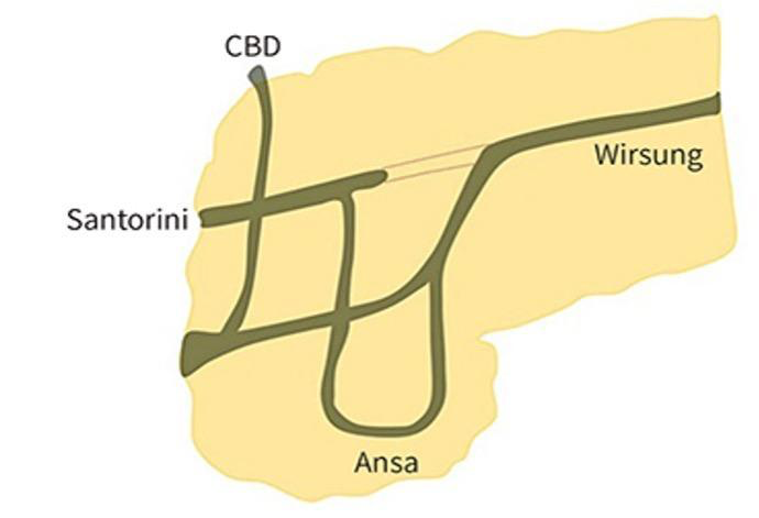
Ansa pancreatica is a rare pancreatic duct variant in which the accessory pancreatic duct forms a looped, S-shaped course before draining through the minor papilla. This altered anatomy may impair pancreatic drainage and has been associated with recurrent acute pancreatitis.
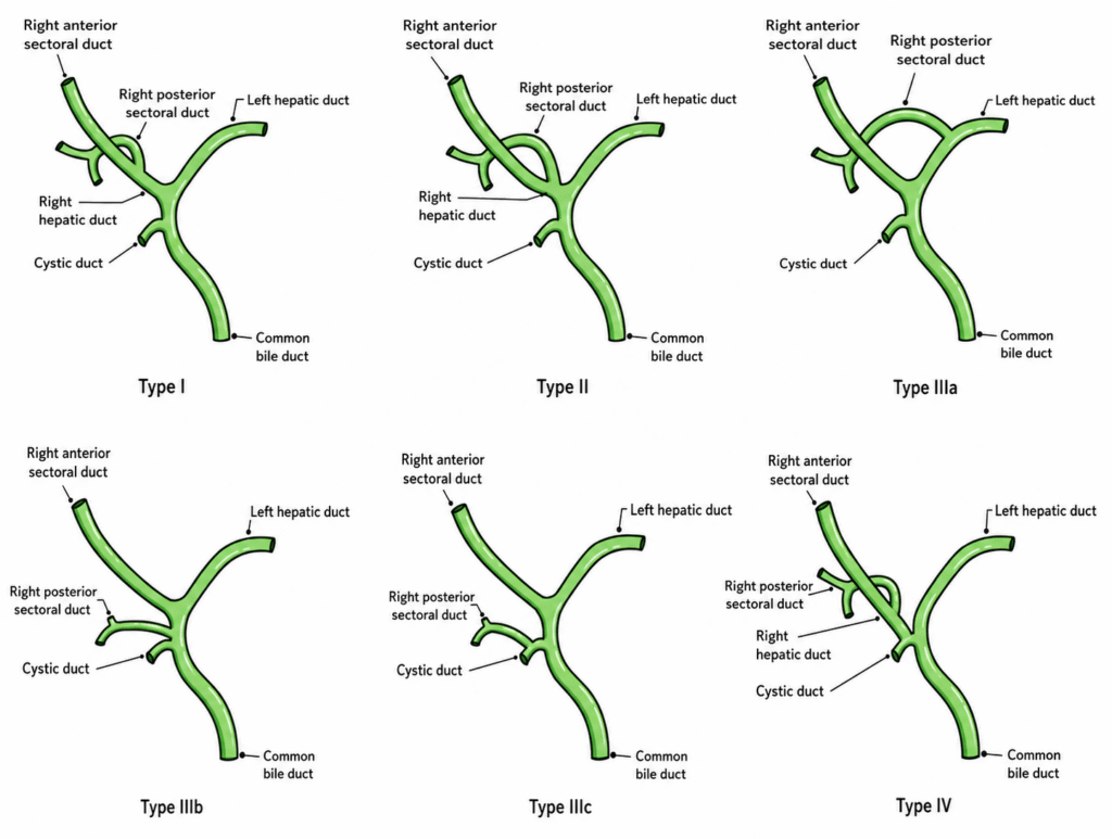
Hilar ductal anatomical variants
Outcomes and alternatives: Emphasize that cannulation may fail in some cases; explain backup plans (repeat ERCP, percutaneous drainage or surgery). Discuss the possibility of staged procedures or additional interventions.
Training and Certification Standards (BSG/JAG)
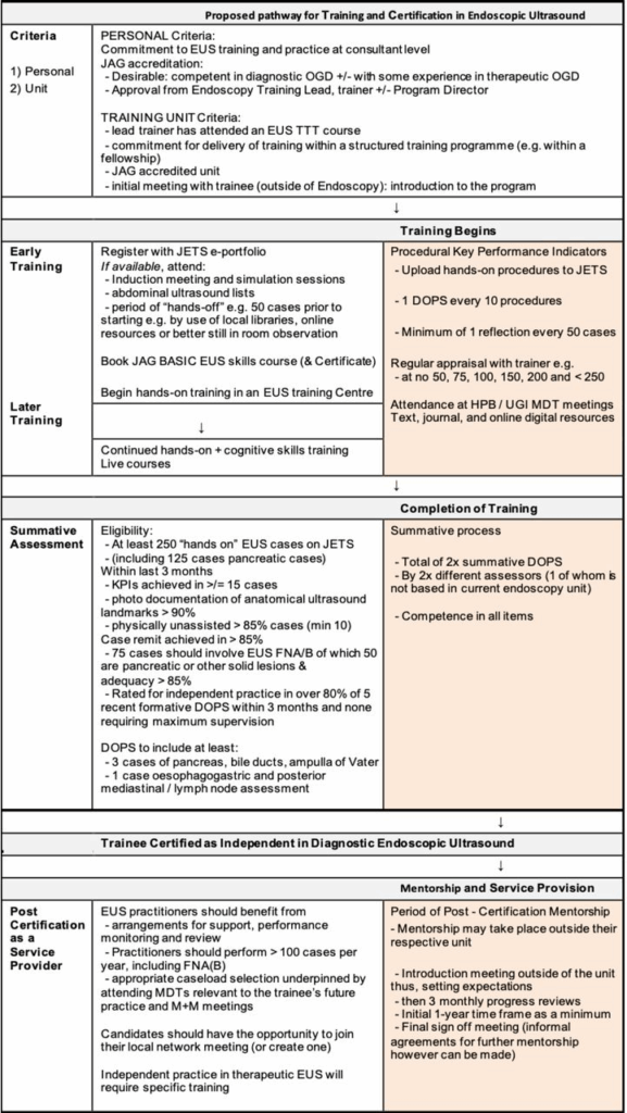
Prerequisites: Trainees must be fully competent in diagnostic OGD (gastroscopy) and ideally in basic upper-GI therapeutic endoscopy before starting ERCP training
JAG requires attendance at a Basic ERCP Skills course (approved centre)
Case volume and skills: UK guidelines recommend a minimum lifetime experience of ~300 supervised ERCPs with at least 30 formative DOPS logged (roughly one per 10 cases). Trainees should achieve key performance targets (for native papillae): ≥80% selective cannulation (last 50 cases) and high success rates in stone clearance and stent placement
Assessment: Progress is tracked via JETS e-portfolio and ERCP DOPS assessments. Certification (for Schutz level 1–2 cases) is granted only after summative assessment by two trainers (different endoscopy units) confirming competence in all domains.
Quality metrics: BSG’s “Way Forward” framework emphasizes training in high-volume ERCP centres. Recommended case-loads are ≥75 ERCPs/year per endoscopist (aspiring to 100+) and ≥150–200 per centre bsg.org.uk. Quality targets include ≥85% cannulation of virgin papillae and ≥75% CBD stone clearance on first ERCP.These benchmarks guide training goals.
Post-certification mentoring: Both BSG and JAG stress ongoing support. Outcomes improve substantially up to 300–800 cases, so newly certified practitioners should have at least 2 years of structured mentorship and regular performance review.
Sources for this ERCP teaching: BSG service standards (2014), JAG training and certification, and BSG consent guidance and update.
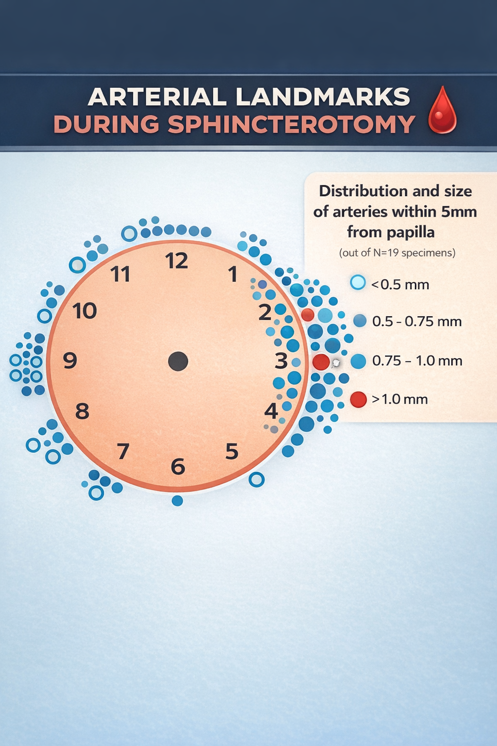
Ampulla and its vascular distribution
Endoscopic ultrasound
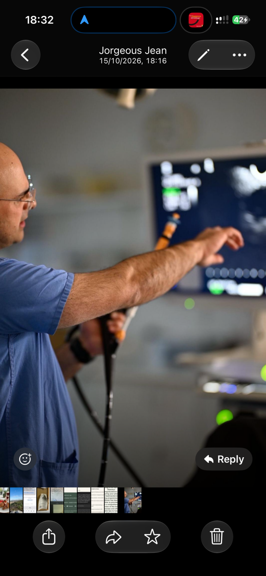
Endoscopic Ultrasound (EUS)
Endoscopic ultrasound (EUS) combines endoscopy with high-frequency ultrasound to delineate GI wall layers and adjacent structures (pancreas, biliary tree, mediastinum). It underpins high-accuracy staging, tissue acquisition (FNA/FNB) and an expanding range of therapeutic procedures.
Linear EUS anatomy: illustrated guide for trainees
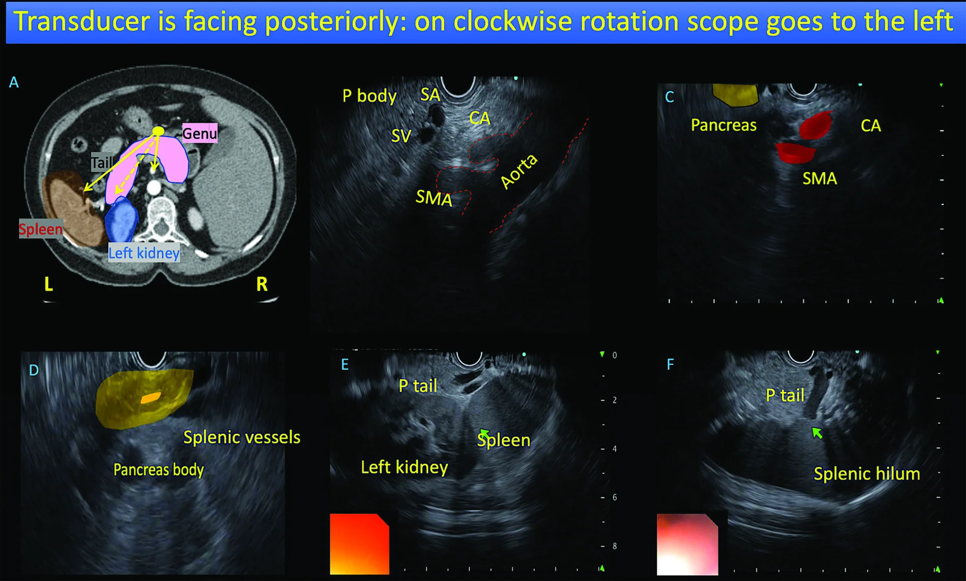
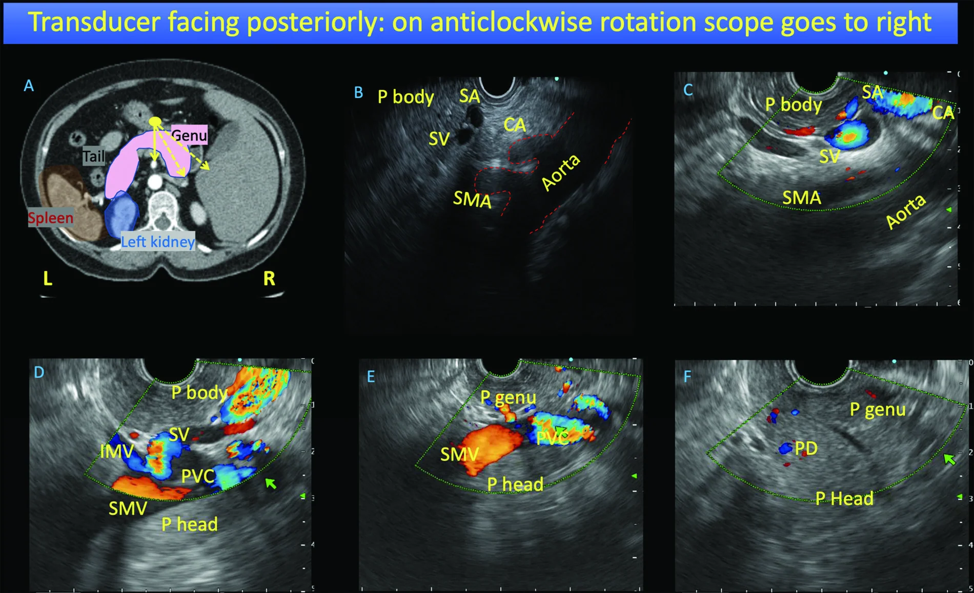
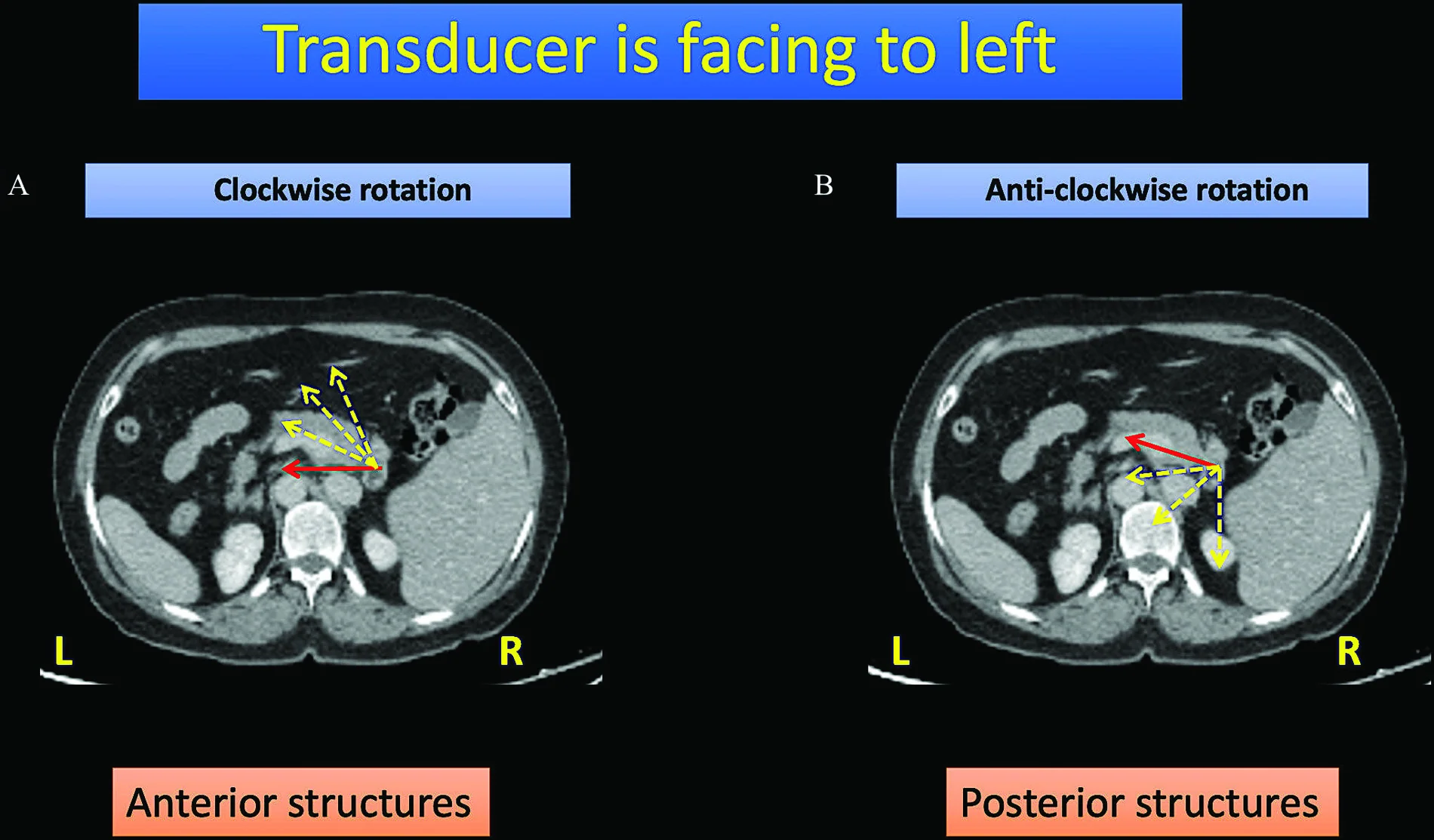
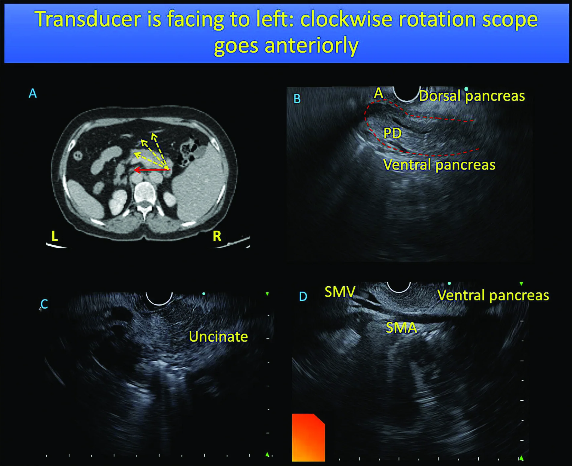
Use these anatomical figures alongside the station-wise technique below. Begin with orientation, trace the pancreatic body and ducts systematically, and optimise the image before interpreting a target.
Orientation and scope rotation
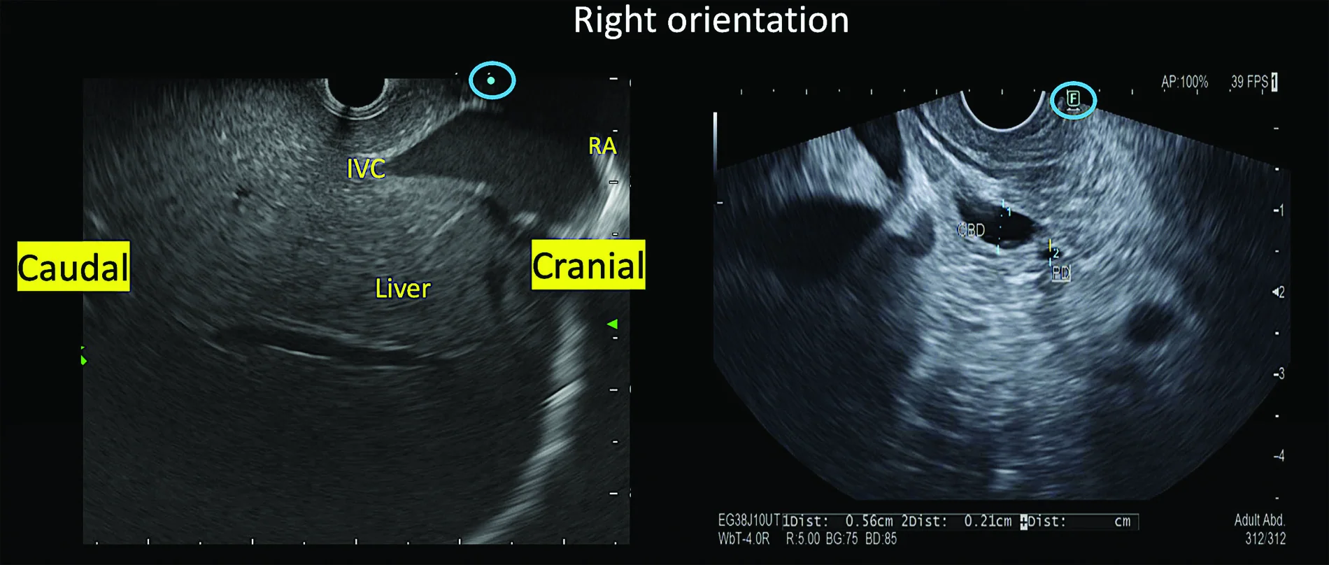
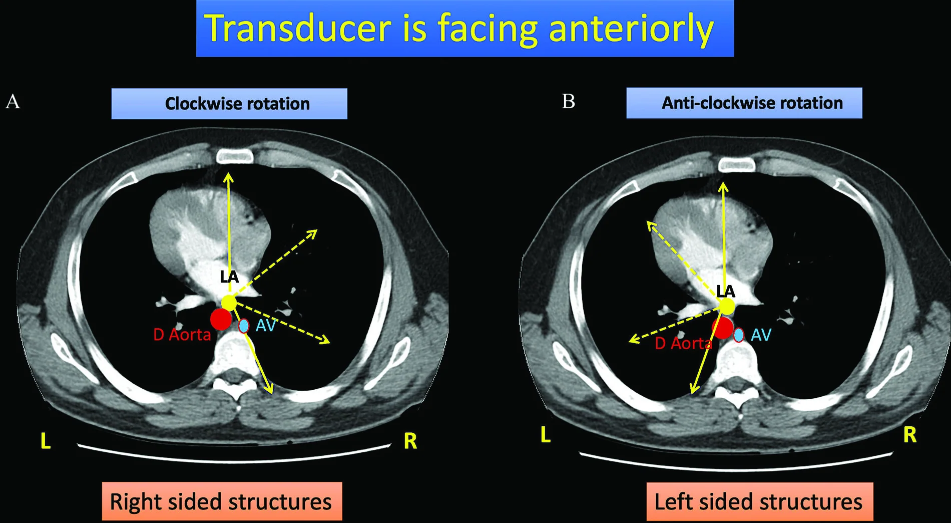
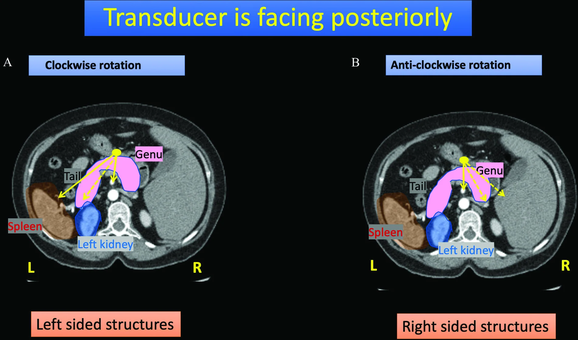
Gastric station: pancreatic body, tail and head
Second part of duodenum (D2)
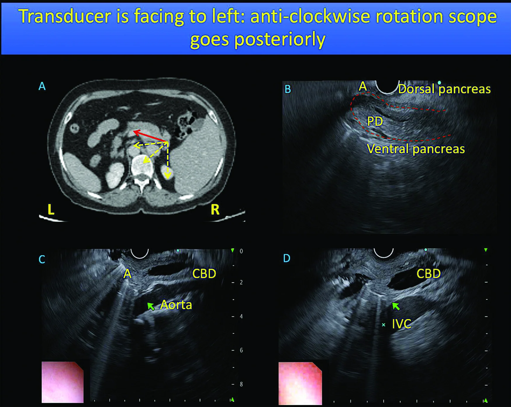
Tracing and centring structures
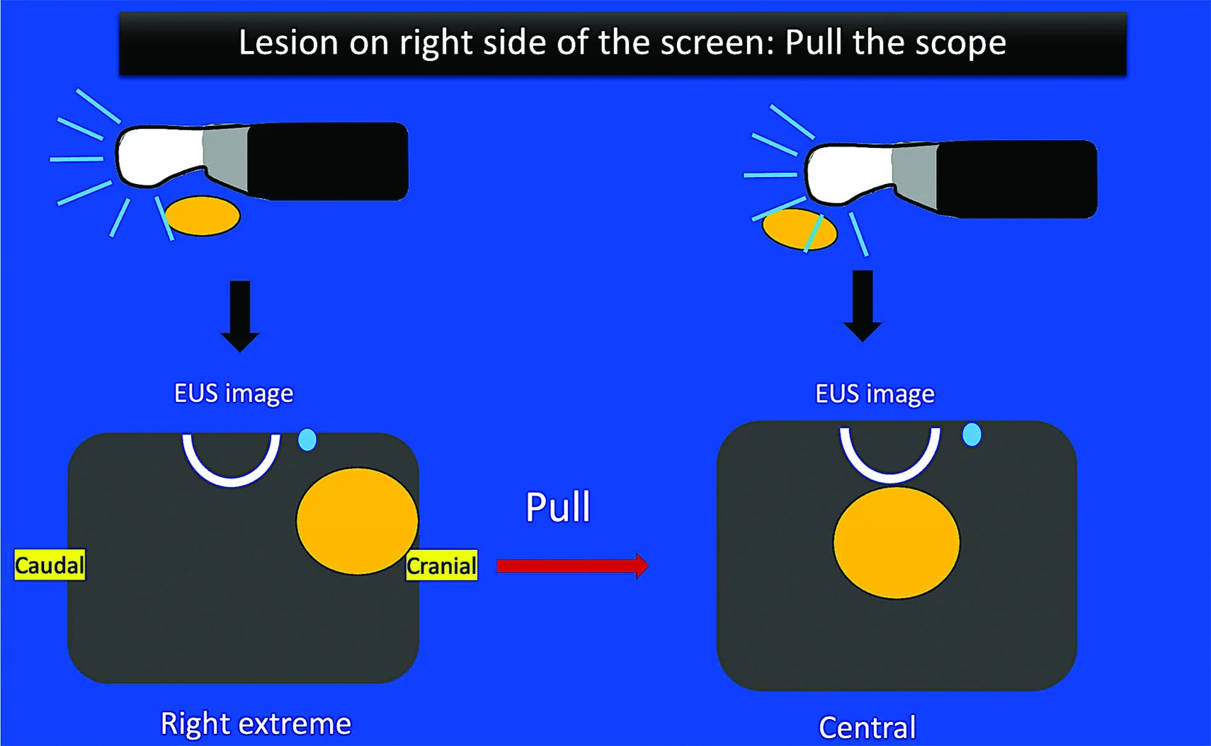
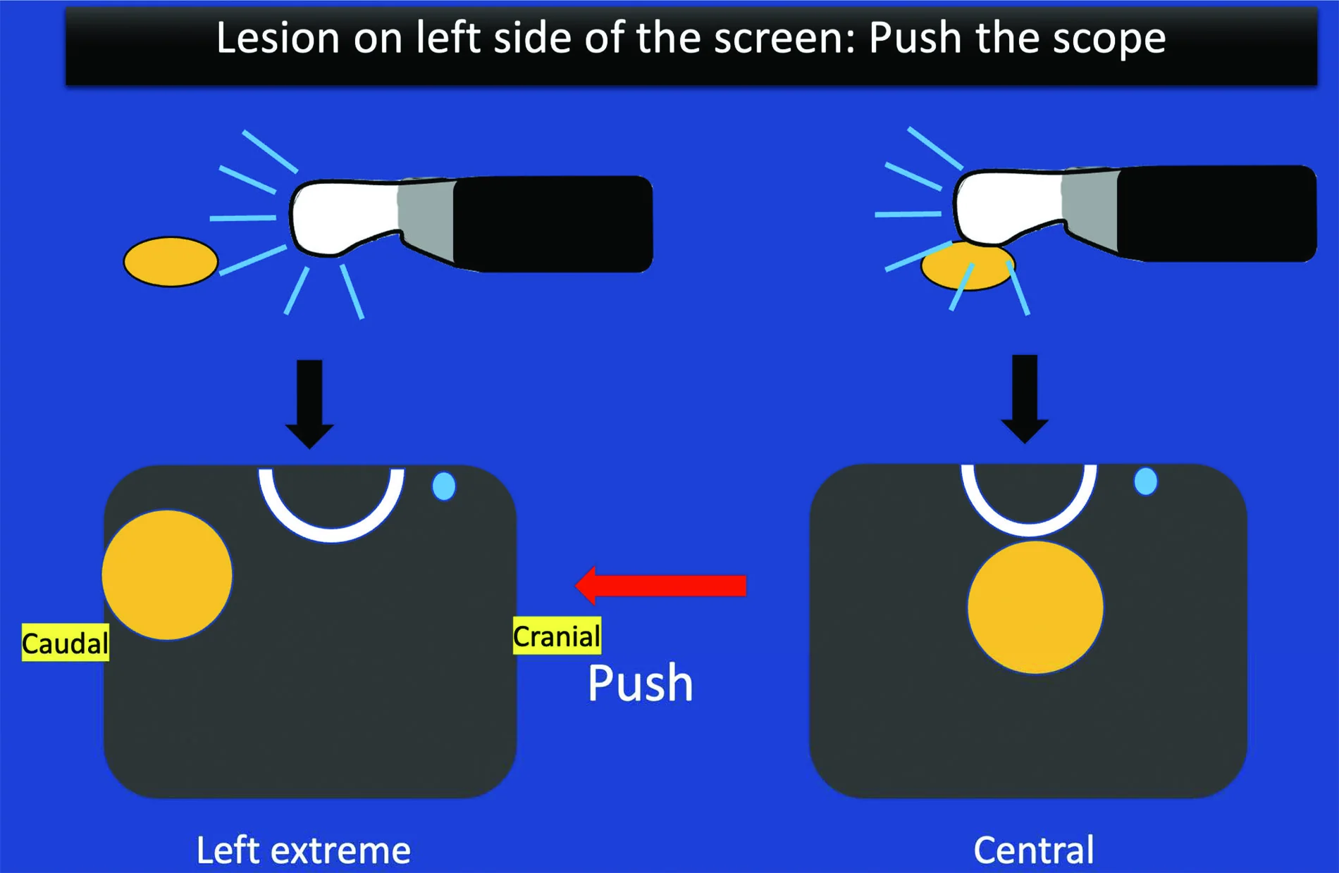
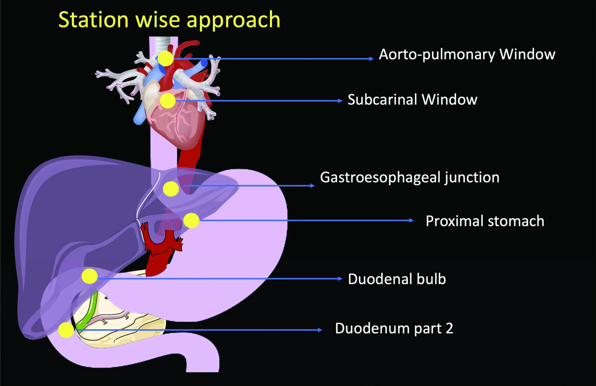
Systematic stations and image optimisation
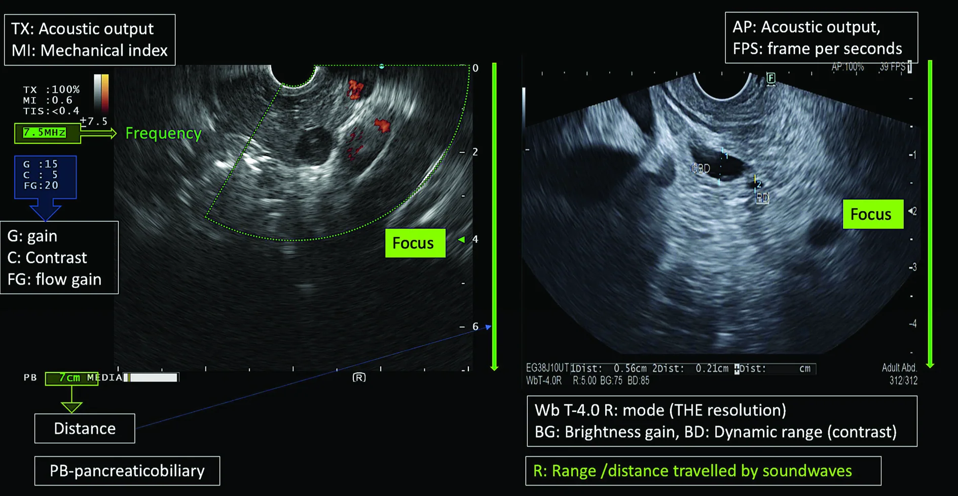
Source and image licensing
Chavan R, Quintini D, Gandhi C, Giri S, Nabi Z, Lakhtakia S, Rajput S. Understanding the Basics of Linear Endoscopic Ultrasound: A Step-by-Step Guide to Anatomy and Orientation. J Dig Endosc. 2025;16:77–90. doi:10.1055/s-0045-1809384.
Original figures © 2025 the authors, reproduced under Creative Commons Attribution 4.0 International (CC BY 4.0). Figures were cropped from the source PDF and converted to WebP for display. Explanatory captions are adapted for trainees. No endorsement by the authors or publisher is implied.
Indications — When to Refer for EUS
1) Oncologic staging
Oesophagus/OGJ: High accuracy for local T/N staging to triage neoadjuvant therapy vs surgery; complementary to CT/PET.
Stomach: Useful for early gastric cancer (endoscopic resectability decisions); superior to CT for local T/N in many series.
Pancreas: Best test for small masses, vascular interface, and pNETs; sensitivity for small tumours >90%.
2) Pancreaticobiliary
Suspected choledocholithiasis: Sensitivity >90%; comparable to MRCP and avoids ERCP risks in intermediate probability.
Pancreatic cysts: Morphology + EUS-FNA (fluid CEA/cytology) to stratify mucinous potential and high-risk features.
Chronic pancreatitis assessment: Structural changes when CT/MRI/US equivocal; correlate clinically to avoid over-calling age-related change.
3) Subepithelial lesions
Define layer of origin, echotexture, margins; EUS-FNA/biopsy for GIST vs leiomyoma and extramural compression.
4) Mediastinal nodes / lung cancer staging
EUS-FNA of posterior/para-oesophageal stations; complementary to EBUS for near-complete minimally invasive mediastinal staging.
5) Therapeutic EUS (where expertise available)
Coeliac plexus block/neurolysis for pancreatic cancer pain.
Transmural pseudocyst drainage (cystogastrostomy) as first-line for suitable collections.
EUS-guided biliary access/drainage after failed ERCP.
Linear EUS of the Pancreas — Station-Wise Technique (Exam Blueprint)
General tips: Optimize coupling (de-air, water instillation), learn the homebase at each station, keep target at 6 o’clock before tracing ducts/vessels, and record labelled clips.
From the stomach (GE junction/body):
Push method: Homebase = aorta → celiac axis; minimal push brings pancreas into view; clockwise to tail (follow splenic vessels), anticlockwise + push to genu/head.
Pull method: From distal body, withdraw to portal vein confluence (PV+SV+SMV); anticlockwise to genu/head; clockwise to tail.
From the duodenal bulb (D1):
Homebase = main portal vein; classic reverse stack sign (CBD–MPV–CHA versus stomach view). Trace CBD and PD towards the ampulla; use water + release pressure to avoid compressing ducts.
From the descending duodenum (D2):
Homebase = aorta. Withdraw with clockwise torque to see head/uncinate, SMV (closest), and the ampulla (confirm CBD and PD joining). If one duct is unseen, fine torque in opposite direction.
Tissue Acquisition — When & How
Solid pancreatic mass: Perform EUS-FNA/FNB; plan needle path to avoid vessels; multiple passes; ROSE if available.
Cystic lesions: Sample only if results change management (CEA/cytology for mucinous/high-risk stratification).
Nodes (mediastinal/peripancreatic): EUS-FNA for diagnosis and staging when imaging is equivocal.
Rapid Reporting Checklist
Indication & pre-test probability (e.g., “intermediate risk CBD stone”, “staging OGJ T1b”)
Stations completed (stomach GE/body; D1; D2) and completeness (tail→ampulla)
Key landmarks (PV confluence; SMA/SMV; CBD/PD; ampulla)
Lesion description (size, layer of origin, borders, echotexture, ductal contact, vascular invasion)
Rosemont features (if CP query): list major/minor and category
Sampling (needle type/gauge; passes; ROSE; immediate complications)
Therapy (if performed) (e.g., pseudocyst drainage; coeliac neurolysis; EUS-BD)
Conclusion/plan (staging impact, MDT actions, surveillance vs resection vs ERCP)
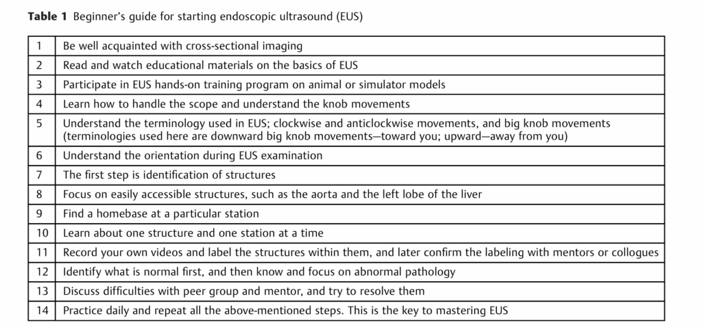
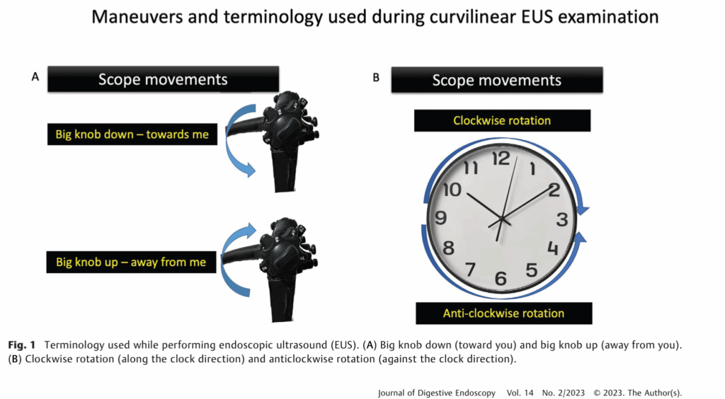
Teaching sources: Reddy and Willert (2009), indications and therapeutic spectrum; Chavan and Rajput (2023), station-wise pancreatic examination. The newer illustrated anatomy section has its own source attribution.
Existing EUS illustrations: Chavan and Rajput (2023). See image attribution and reuse status.
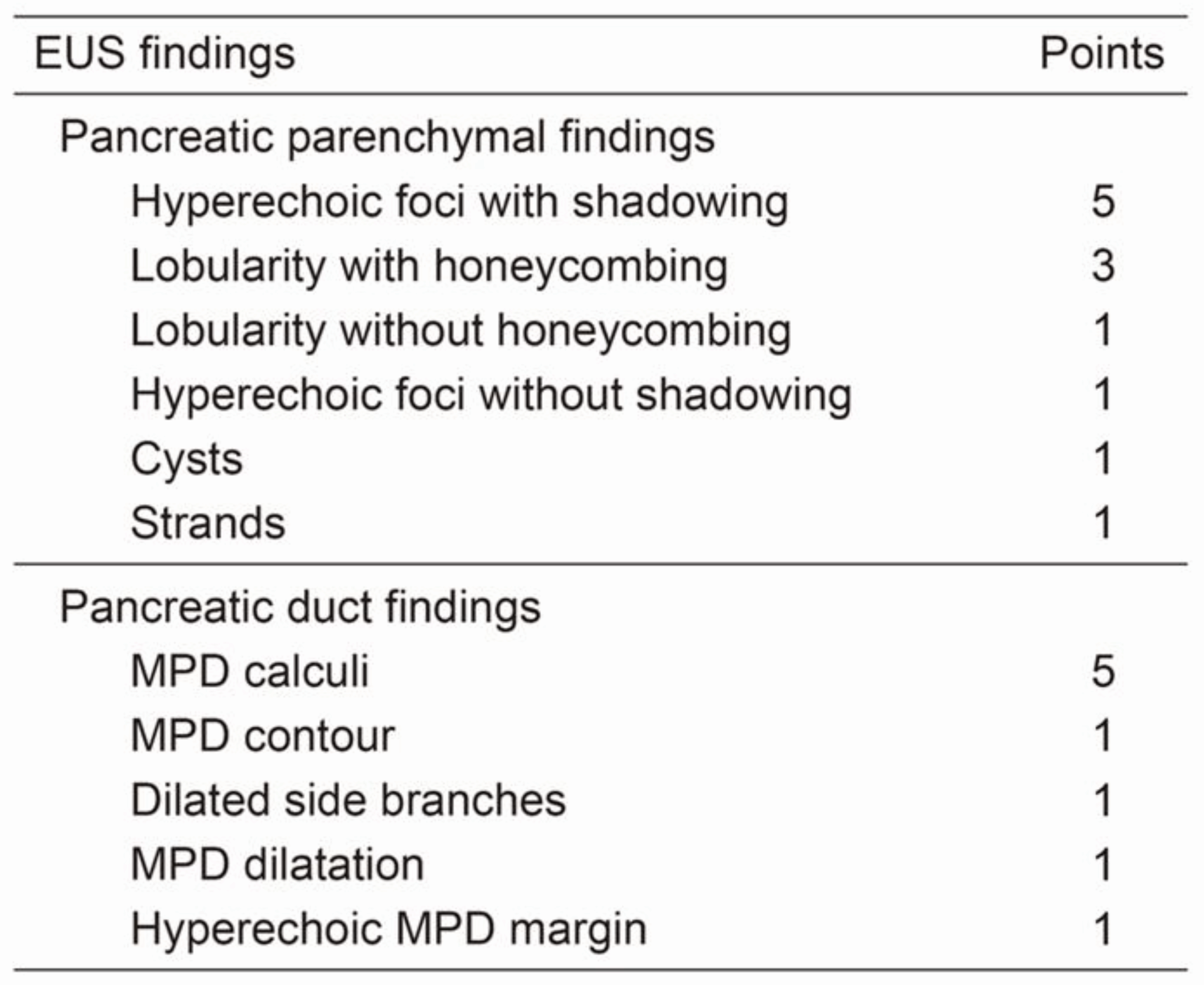
Rosemont criteria (2009)
Major features:
Major A: Hyperechoic foci with acoustic shadowing (calcifications)
Major B: Lobularity with honeycombing
Minor features:
Hyperechoic strands; hyperechoic foci (no shadowing); lobularity without honeycombing; cysts; MPD dilatation; irregular MPD contour; side-branch dilatation; hyperechoic duct wall.
Categories:
Consistent with CP: Major A + Major B or Major A + ≥3 minor or Major B + ≥3 minor
Suggestive: Major A/B + <3 minor or ≥5 minor
Indeterminate: 3–4 minor, no major
Normal: <3 minor, no major
Use in clinical context (history, pain phenotype, pancreatic function, calcific burden) and with cross-sectional imaging; avoid over-calling age-related changes.
EUS-guided therapies
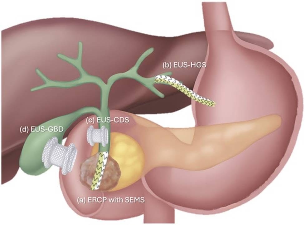
Recent advances in therapeutic EUS mean that endoscopy can now be used not only to diagnose disease, but also to create internal drainage pathways when standard ERCP is difficult or unsuccessful. Lumen-apposing metal stents (LAMS) have played a major role in this progress because they allow secure, transluminal drainage in a single procedure. In specialist centres, EUS-guided choledochoduodenostomy (EUS-CDS/CDD) can be used to drain the common bile duct into the duodenum, while EUS-guided hepaticogastrostomy (EUS-HGS) can provide biliary drainage through the stomach when access from the duodenum is not suitable. EUS has also expanded into other therapeutic areas, including gallbladder drainage, pancreatic fluid collection drainage, and gastroenterostomy. These are advanced procedures that require expert training, careful setup, and close teamwork between the endoscopist, nurses, and radiographer.
References and image credits
Clinical evidence and guidance
- ERCP service standards. Wilkinson M, et al. ERCP – The Way Forward: A Standards Framework. British Society of Gastroenterology; June 2014. Source for the service-volume and performance standards discussed above.
- ERCP training. Joint Advisory Group on GI Endoscopy. JAG training pathway and certification standards for ERCP. See also the current JETS certification resources.
- Consent. Everett SM, Griffiths H, Nandasoma U, et al. Guideline for obtaining valid consent for gastrointestinal endoscopy procedures. Gut. 2016;65:1585–1601. Original guideline; BSG guideline and February 2023 update.
- Ampullary vascular anatomy. Mirjalili SA, Stringer MD. The arterial supply of the major duodenal papilla and its relevance to endoscopic sphincterotomy. Endoscopy. 2011;43:307–311. Original publication.
- Cannulation and sphincterotomy. Testoni PA, Mariani A, Aabakken L, et al. Papillary cannulation and sphincterotomy techniques at ERCP: ESGE Clinical Guideline. Endoscopy. 2016;48:657–683. Original guideline (PDF).
- EUS indications. Reddy Y, Willert RP. Endoscopic ultrasound: what is it and when should it be used? Clinical Medicine. 2009;9:539–543. Original review. This is the historical source review used for the indications summary.
- Station-wise linear EUS. Chavan R, Rajput S. Pictorial Essay of Linear Endoscopic Ultrasound Examination of Pancreas Anatomy. Journal of Digestive Endoscopy. 2023. Original publication.
- Illustrated linear EUS anatomy. The full Chavan et al. (2025) citation and figure-specific licence information accompany the 12 illustrated anatomy figures.
- Rosemont classification. Catalano MF, Sahai A, Levy M, et al. EUS-based criteria for the diagnosis of chronic pancreatitis: the Rosemont classification. Gastrointestinal Endoscopy. 2009;69:1251–1261. Original consensus publication.
- Early chronic pancreatitis: separate diagnostic criteria. Kashima K, Yamamiya A, Abe Y, et al. Proposal and Validation of New Diagnostic Criteria for Diagnostic Weights of Endoultrasonographic Findings for Early Chronic Pancreatitis. Journal of Clinical Medicine. 2023;12:5320. Original publication. This paper is distinct from the original Rosemont consensus.
- Therapeutic EUS and palliation. Gainey CS, Baron TH. Endoscopic Approach to Palliation of Pancreatic Cancer. The American Journal of Gastroenterology. Publication record.
Image attribution and reuse status
Image credits and reuse conditions are listed separately from the clinical evidence above. A clinical citation does not itself establish permission to reproduce an image.
- Chavan et al. (2025), 12 anatomy figures: CC BY 4.0 attribution, source link and crop/WebP adaptation notes are retained with every figure and in the anatomy section. The publisher identifies the article as CC BY 4.0.
- Ansa pancreatica: Galzerano C, et al. Acute pancreatitis in a patient with “ansa pancreatica”. Eurorad case 18292; 3 October 2023. DOI: 10.35100/eurorad/case.18292. CC BY-NC-SA 4.0; the non-commercial and share-alike conditions apply.
- Existing beginner EUS and manoeuvre illustrations: attributed on this page to Chavan and Rajput (2023). Figure-level correspondence and reuse permissions have not been independently established in this audit.
- Table displayed alongside Rosemont teaching: the existing credit identifies Kashima et al. (2023), published under CC BY 4.0. The exact table correspondence and any third-party exceptions require confirmation against the publisher version; this is not a verified attribution to the original Rosemont paper.
- Regional pancreatic cyst pathway, hilar anatomy variants, ampullary blood-supply illustration, EUS training image and therapeutic drainage illustration: no independently verified reuse permission was established. The ampullary evidence sources are listed above; the therapeutic illustration has an existing Gainey–Baron source credit. Evidence references are not image licences.
- EUS-guided biopsy photograph: supplied for this website by its owner; the existing caption is preserved. No third-party open licence is asserted.