
Clinical resources / Hepatology
Liver medicine.
A clearer view.
Clinical summaries, investigation guidance and practical learning resources for your hepatology practice.
All treatment guidelines are given for information only – please always follow local guidelines and formularies.
Introduction
Patients often require checking of their liver function tests for a number of reasons both in the inpatient and outpatient settings. Liver blood or function tests (LFTs) are an inexpensive way of commencing investigations in those suspected of liver disease. Those patients with abnormal liver blood tests should be considered for further investigation with a liver aetiology screen. LFTs can also be used in the monitoring of potential adverse effects of drugs on the liver such as statins, and for the investigation of the generally unwell patient.
When interpreted in isolation, hepatobiliary enzymes can provide information on the level of ongoing injury, whereas bilirubin, albumin and INR convey information on liver function, with platelets conveying information on the level of fibrosis.

Clinical History
A full clinical history can is essential in determining a potential cause for LFT derangement.
Age
Ethnicity/country of birth (to explore possible risk of hepatitis B or C)
Specific symptoms (jaundice, abdominal pain, weight loss, pruritus, etc)
Comorbidities
Drug history (prescribed, over the counter, herbal, injecting drug use, illicit)
Travel history
Occupational exposure
Alcohol history (current and past intake in average units per week)
Features of the metabolic syndrome (central obesity, hypertension, diabetes/insulin resistance and dyslipidaemia)
Family history
BSG / BASL quick reference
Cirrhosis management
Ward and clinic guidance, including SBP and the decompensated cirrhosis admission bundle.
First six hours: bundle essentials
- Start the admission bundle within six hours. Record NEWS and suspected precipitants.
- Sepsis screen: blood cultures, urine testing/culture and CXR. Request abdominal ultrasound with hepatic and portal vein Doppler.
- Perform an ascitic tap promptly. Do not delay to correct cirrhosis-related coagulopathy.
- Assess VTE prevention. The bundle recommends prophylaxis unless active bleeding or platelets <50 × 10⁹/L, with individual contraindications considered.
- Ongoing alcohol use: IV thiamine/B vitamins, withdrawal assessment using CIWA or GMAWS, and monitor for refeeding syndrome under local protocols.
- Refer early to hepatology and dietetics. Reassess AKI at six hours and escalate deterioration to HDU/ICU.
01Routine care
- Treat the underlying cause. Support alcohol abstinence.
- Review symptoms, blood results, disease severity, medicines, nutrition and frailty.
- Arrange six-monthly ultrasound and AFP for HCC surveillance when treatment could benefit the patient.
- Assess portal hypertension, vaccination needs and fracture risk.
02Acute decompensation
- Look for infection, bleeding, alcohol-related injury, dehydration and medication effects.
- Check FBC, renal/liver profile, INR, CRP, glucose, lactate, magnesium and phosphate. Obtain cultures and appropriate imaging.
- Tap ascites promptly on admission. Send neutrophil count, culture, protein and albumin. Inoculate blood-culture bottles at the bedside.
- Escalate shock, respiratory failure, severe encephalopathy or worsening AKI.
03Ascites
- First moderate presentation: spironolactone 100 mg daily. Recurrent severe ascites or need for faster diuresis: add furosemide 40 mg daily. Titrate with weight, renal function and electrolyte monitoring.
- Tense ascites: therapeutic paracentesis. After removing >5 L, give albumin 8 g per litre of total ascites removed. Consider albumin after smaller drains in ACLF or high AKI risk.
- Recurrent/refractory ascites: discuss TIPSS and transplant assessment.
04Spontaneous bacterial peritonitis
- Start empirical IV antibiotics. Follow local policy, acquisition setting and resistance risk. Do not await cultures.
- Albumin: 1.5 g/kg within six hours, then 1 g/kg on day 3. Use estimated dry weight and monitor for fluid overload.
- Consider a repeat tap at 48 hours if response is inadequate or secondary peritonitis is suspected. Investigate an intra-abdominal source.
- After recovery, consider secondary antibiotic prophylaxis with hepatology/microbiology. Individualise primary prophylaxis.
Albumin eligibility: The 2025 admission guidance recommends albumin for SBP. The 2021 ascites recommendation specifically identifies patients with increased/rising creatinine.
05Portal hypertension & varices
- Compensated cirrhosis with clinically significant portal hypertension: consider carvedilol 6.25 mg daily, increasing to 12.5 mg/day if tolerated.
- Check contraindications, BP and renal function. Individualise use in decompensated disease.
- If beta-blockers are unsuitable, arrange OGD and band high-risk oesophageal varices.
- Reduce/withhold beta-blockers with persistent systolic BP <90 mmHg or acute/progressive renal dysfunction.
06Suspected variceal bleeding
- Resuscitate, assess the airway and contact the GI bleeding team.
- Start IV vasoactive treatment and antibiotics. Use restrictive transfusion, adjusted for shock and comorbidity. Suspend beta-blockers during terlipressin/octreotide treatment. Do not routinely correct INR with blood products.
- OGD after resuscitation, ideally within 12 hours. BSG permits up to 24 hours in haemodynamically stable patients depending on resources. Band bleeding oesophageal varices.
- Uncontrolled bleeding: urgent rescue TIPSS discussion, with a stent or balloon bridge where appropriate.
- Secondary prevention: beta-blocker plus repeat banding unless a patent salvage/rescue TIPSS is in place. Gastric/ectopic varices need specialist planning.
BSG June 2026: routine pre-emptive TIPSS is not recommended outside research settings.
07Hepatic encephalopathy
- Treat infection, bleeding, constipation, dehydration, electrolyte disturbance and sedative effects.
- Lactulose 20–30 mL up to four times daily, titrated to 2–3 soft stools daily. Avoid excessive diarrhoea.
- Add rifaximin 550 mg twice daily for recurrent overt episodes.
- Avoid protein restriction. Severe encephalopathy needs urgent airway assessment.
08AKI & suspected HRS
- Stop diuretics and nephrotoxic medicines. Assess infection, bleeding and volume status.
- If hypovolaemic, give crystalloid in 250 mL boluses with repeated assessment. Monitor urine output and creatinine.
- Persistent AKI: urgent specialist assessment for alternative causes and HRS-directed treatment. Avoid indiscriminate fluid loading.
09Nutrition & longer-term planning
- Arrange dietetic input. Encourage frequent meals, a bedtime snack and adequate protein.
- Discuss transplant assessment after decompensation, particularly with recurrent complications.
- Include symptom control, patient priorities and advance care planning alongside active treatment.
BSG / BASL / SAM admission bundle
Official two-page form · Version 2.0 · 12 January 2025
Open or download the original bundle (PDF)
Source clarification: the original form prints Hb “7–8 g/L”. Read this as 70–80 g/L (7–8 g/dL), individualised for bleeding and comorbidity. Clinical dehydration warrants assessment and treatment but alone does not meet KDIGO AKI diagnostic criteria.
Hepatitis B Management
Phases of Infection:
Immune Tolerant Phase: Common in younger patients infected perinatally or in childhood. Characterized by high HBV DNA (>1×10⁷ IU/mL) and normal ALT. Risk of progression to fibrosis is low, but patients should be monitored every 6–12 months with ALT, HBV DNA, HBsAg, and HBeAg status. Fibroscan should be performed every 2–3 years
Immune Active Phase: Patients exhibit elevated ALT and HBV DNA (>2000 IU/mL). Immediate treatment with Tenofovir or Entecavir is recommended to suppress viral replication and reduce liver inflammation. Monitoring involves 3–6 monthly checks during active treatment, then 6–12 months once stable
Inactive Carrier: Patients are HBeAg negative, with low or undetectable HBV DNA. Monitor for ALT flares and reactivation every 6–12 months. Treat if reactivation occurs
Cirrhotic Patients:All require antiviral therapy irrespective of HBV DNA levels to prevent further decompensation and HCC development.Perform 6-monthly ultrasound and AFP for HCC surveillance and refer for varices screening if Fibroscan >20 kPa or platelets <150
Special Situations:
- Hepatitis B Reactivation: Treat reactivation aggressively if ALT is elevated and HBV DNA >2000 IU/mL.
- Pregnancy: Use Tenofovir from 32 weeks if maternal HBV DNA >10⁶ IU/mL to prevent vertical transmission. Infants require HBV vaccine and immunoglobulin within 24 hours of birth
Autoimmune Hepatitis
Treatment protocols often vary from trust to trust – here is a summary of the NUTH ( Newcastle guidelines)
Induction Therapy:Start with Prednisolone 40 mg daily. Taper weekly by 5 mg to reach a maintenance dose of 10 mg, adjusting based on therapeutic response (normalization of ALT and IgG). Consider Budesonide in non-cirrhotic patients for fewer systemic side effects.
Maintenance Therapy:Alternatives include Mycophenolate Mofetil for Azathioprine intolerance or Tacrolimus for refractory cases.Azathioprine is the first-line option (1 mg/kg/day). It is contraindicated in acute presentations with jaundice and requires TPMT testing before initiation
Monitoring: Patients in remission need 6-monthly clinic reviews and blood tests every 3 months. Cirrhotic patients require additional HCC surveillance with ultrasounds every 6 months.
Prognosis: Withdrawal of immunosuppression may be considered after 2–3 years of sustained biochemical remission, although relapse rates are high
Primary Biliary Cholangitis
First Line: Ursodeoxycholic acid (UDCA) 13–15 mg/kg/day. Evaluate response after 12 months using ALP and bilirubin thresholds (e.g., POISE criteria: ALP >1.67x ULN indicates incomplete response)
Second Line: Obeticholic acid or Bezafibrate for patients who do not respond or are intolerant to UDCA. Start at low doses (e.g., Obeticholic acid 5 mg/day) with close monitoring
Surveillance:
Cirrhotic patients need biannual imaging and AFP for HCC surveillance. Bone density assessment is recommended every 5 years due to the risk of osteoporosis
Management of Symptoms:
- Itch: Options include cholestyramine, rifampicin, or gabapentin. Regular iron studies are advised as iron deficiency may exacerbate symptoms
- Fatigue: No proven therapies; screen for and treat contributing factors such as thyroid dysfunction or vitamin deficiencies.
Primary Sclerosing Cholangitis
Therapy:
While there are no licensed long-term therapies, UDCA (13–15 mg/kg/day) may improve liver biochemistry but does not alter long-term outcomes.Regular Fibroscan helps monitor disease progression
Surveillance:
Stable PSC without cirrhosis: yearly liver and gallbladder ultrasound, with MRCP every 2–3 years according to the individual hepatology plan. Arrange earlier MRI/MRCP if symptoms, liver blood tests or ultrasound findings raise concern.
If cirrhosis is present: continue 6-monthly ultrasound for HCC surveillance.
Patients with concomitant IBD require annual colonoscopy with biopsies to screen for colorectal cancer.
EASL PSC guidance (2022) suggests at least yearly ultrasound and/or MRI/MRCP for large-duct PSC. A fixed 2–3-year MRCP interval is an individualised schedule, not a universal EASL recommendation.
Acute Liver Failure
Referral Criteria:
Transfer to a specialized center if encephalopathy, INR >2, or bilirubin >300 μmol/L is present. Early intubation and ventilation should be considered for safe transport. Discussion with teriatry center when PT is increasing and approaching 50.
Management:Initiate N-Acetylcysteine (NAC) for both paracetamol and non-paracetamol etiologies.Correct coagulopathy judiciously; avoid routine INR correction unless invasive procedures are planned. Regularly monitor blood glucose to prevent hypoglycemia. Sodium levels should be kept at 140–145 mmol/L to minimize the risk of cerebral edema.
Chronic liver disease
On a ward round
When managing patients with decompensated liver disease, it is essential to identify and address potential triggers such as infections, thrombosis, constipation, or bleeding.
Nutritional needs should be assessed promptly, with dietitian involvement prioritized in cases of electrolyte imbalances or sarcopenia.
Specialist support, including alcohol specialist nurses or drug addiction teams, should be engaged as needed.
For patients with cirrhosis, evaluate for varices via OGD or liver stiffness exceeding 20 kPa and initiate low-dose carvedilol if blood pressure permits, adjusting the dose as required.
Ensure that six-monthly ultrasound surveillance for hepatocellular carcinoma (HCC) is up to date and perform DEXA scans every three years to monitor bone health.
In patients with a history of encephalopathy, secondary prophylaxis with lactulose and rifaximin should be implemented. This comprehensive approach helps address the multifaceted needs of these patients effectively.
BSG useful articles
All open access recommended selected articles
NAFLD: Comprehensive guidelines for managing non-alcoholic fatty liver disease.
https://www.bsg.org.uk/clinical-resource/quality-standards-for-the-management
Compensated Cirrhosis Management: Guidance on outpatient care for patients with compensated cirrhosis.
https://www.bsg.org.uk/clinical-resource/bsg-guidance-outpatient-management-of-cirrhosis
Decompensated Cirrhosis Management: Recommendations for managing decompensated cirrhosis, including key complications.
https://www.bsg.org.uk/clinical-resource/bsg-guidance-outpatient-management-of-cirrhosis-2
Cirrhosis Special Circumstances: Insights into managing cirrhosis in special scenarios like surgery, pregnancy, and travel.
https://www.bsg.org.uk/clinical-resource/bsg-guidance-outpatient-management-of-cirrhosis-3
Wilson disease
BSG useful articles
All open access recommended selected articles
Overview
Wilson disease is an autosomal recessive disorder that impairs copper metabolism, predominantly affecting the liver and nervous system. Symptoms usually appear between the ages of 5 and 35, often leading to serious complications if untreated.
Diagnosis
Diagnosis requires a combination of clinical findings and laboratory tests. The presence of Kayser-Fleischer rings alongside low serum ceruloplasmin levels (less than 100 mg/L) is highly suggestive of Wilson disease. If Kayser-Fleischer rings are absent, the diagnosis is confirmed by meeting at least two of the following criteria: low serum ceruloplasmin, urinary copper excretion greater than 1.6 µmol per 24 hours, a positive ATP7B gene mutation, or hepatic copper levels exceeding 250 µg per gram of dry weight.
Indications for Liver Transplantation
Liver transplantation is necessary in cases of acute liver failure or decompensated chronic liver disease resulting from Wilson disease.
Treatment
Management of Wilson disease focuses on reducing copper accumulation. Copper-chelating agents such as penicillamine or trientine are first-line therapies. Zinc acetate serves as a second-line option, functioning to reduce copper absorption in the gastrointestinal tract.
Screening
Genetic and biochemical screening of family members is critical due to the hereditary nature of the disease. The probability of siblings being homozygous for Wilson disease is 25%, while the risk for offspring is approximately 0.5%.
Monitoring
The effectiveness of treatment is monitored through urinary copper measurements to ensure proper copper excretion and control of the disease.
This approach to diagnosis, treatment, and monitoring is consistent with the European Association for the Study of the Liver (EASL) 2012 guidelines.
References:
Newcastle upon Tyne Hospitals NHS Foundation Trust. NUTH Hepatitis B Guidelines. Version 11.0, February 2024.
Newcastle upon Tyne Hospitals NHS Foundation Trust. Autoimmune Liver Disease Clinic Guidelines. August 2024.
Newcastle upon Tyne Hospitals NHS Foundation Trust. Autoimmune Liver Disease Clinic Guidelines. August 2024.
Newcastle upon Tyne Hospitals NHS Foundation Trust. Autoimmune Liver Disease Clinic Guidelines. August 2024.
Newcastle upon Tyne Hospitals NHS Foundation Trust. Guidelines for the Management of Acute Liver Failure. Version 3, Effective from March 9, 2024.
Newcastle upon Tyne Hospitals NHS Foundation Trust. Liver Transplant Unit Guidelines. Version 6, Effective from March 11, 2024.
Newcastle upon Tyne Hospitals NHS Foundation Trust. Liver Unit Crib Sheet. 2024.
FibroScan: a practical interpretation guide
Two different measurements: liver stiffness (kPa) helps assess fibrosis; CAP (dB/m) assesses liver fat. Neither is a direct measure of liver function. Use the clinical history, blood tests and imaging alongside the scan.
What do F0–F4 mean?
| Stage | Meaning |
|---|---|
| F0 | No fibrosis |
| F1 | Mild fibrosis |
| F2 | Moderate / significant fibrosis |
| F3 | Advanced / bridging fibrosis, without established cirrhosis |
| F4 | Cirrhosis |
These are tissue-based stages. FibroScan values overlap: there is no universal kPa-to-F-stage conversion. [2]
Liver stiffness in MASLD
Advanced fibrosis unlikely; mild fibrosis remains possible.
Indeterminate: further assessment.
Supports advanced fibrosis; specialist assessment. Not proof of cirrhosis.
MASLD-specific VCTE thresholds, not universal cut-offs. [1, Table 6]
A practical MASLD pathway
- FIB-4 first: <1.3 → reassess in 1–3 years; 1.3–2.67 → VCTE/ELF; >2.67 → hepatology referral.
- Age adjustment: above 65, use 2.0 as the lower cut-off. FIB-4 performs poorly below 35.
- Intermediate FIB-4: alternatively, intensify metabolic management for one year, then perform elastography if still elevated. VCTE ≥8 kPa warrants further evaluation. [1, Fig. 2]
CAP: liver fat, not fibrosis
| Suggested CAP threshold | Steatosis grade |
|---|---|
| 248 dB/m | S1 |
| 268 dB/m | S2 |
| 280 dB/m | S3 |
These are approximate thresholds, not precise fat percentages or rigid bands. CAP does not diagnose MASH or stage fibrosis. [1, Table 6]
Check reliability before acting
- Fast for at least 3 hours. Check probe selection and the report’s measurement-quality indicators.
- Inflammation, cholestasis, congestion and recent food intake may elevate stiffness independently of fibrosis.
- VCTE, ultrasound shear-wave elastography and MR elastography have different validated thresholds; do not convert between them using a fixed offset.
- If results are unreliable or disagree with the clinical picture, repeat under appropriate conditions and/or use another validated test. Specialist review and sometimes biopsy are needed when uncertainty would change management. [2]
Portal hypertension: a separate assessment
For compensated advanced chronic liver disease; these thresholds do not assign a fibrosis stage.
- LSM ≤15 kPa and platelets ≥150 × 109/L: can rule out clinically significant portal hypertension (CSPH).
- LSM ≥25 kPa: can rule in CSPH in viral/alcohol-related disease and non-obese MASLD (BMI <30). Do not apply this rule-in threshold uncritically in obesity.
- Varices assessment: if unsuitable for non-selective beta-blockers, screen by endoscopy when LSM ≥20 kPa or platelets ≤150 × 109/L. Those already receiving beta-blockers to prevent decompensation generally do not need screening endoscopy solely to detect varices. [3]
Original scoring chart — historical reference
The chart’s disease-specific bands are illustrative, not current universal EASL cut-offs. Use the guide above for MASLD. On mobile, swipe across the chart or tap to open full-size.
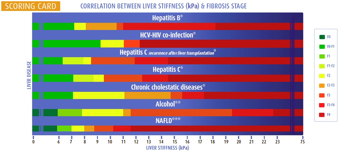
Source: Dr Douglas Samuel’s Fibroscan resource. The original chart uses the older term NAFLD.
References
- EASL–EASD–EASO MASLD guideline (2024): Figure 2 and Table 6.
- EASL non-invasive liver tests guideline (2021).
- Baveno VII consensus (2022), also informing EASL MASLD guidance.
Educational summary • Updated 28 September 2026 • Follow local hepatology pathways.
Continue learning